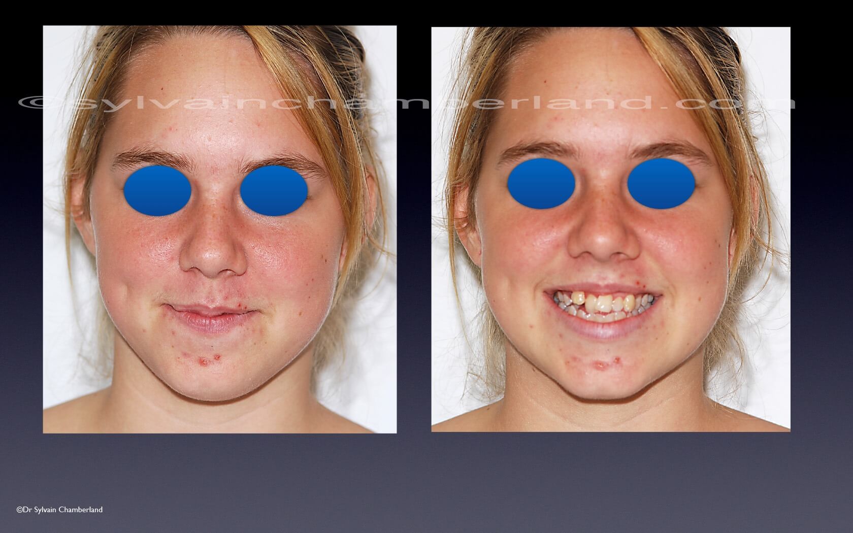
Class I. Coronal destruction of upper 1st molar #16
Severe lack of space in maxilla. Extensive decay on 1st molar. Poor prognosis of this 1st molar (#16).
Palatal ectopy of the right upper lateral incisor.
Upper mid-arch deviation to the right.
Anterior protrusion
Dentoalveolar protrusion
Class III relationship of bony bases.
What is the best treatment plan for this patient?
Update, Sept. 8, 2019
Thanks to everyone who participated and gave their opinion.
There was one unavoidable element. The 1st molar had a guarded prognosis and had to be extracted.
The 2nd element is the planning of dental movements.
The “Visual treatment Objective” slide explains how the teeth should be positioned. It’s important to visualize the retreatment of the lower incisors in order to achieve a positive overjet. Because of the lack of space in the maxilla, the upper incisors will recede little or not at all.
The use of a compressed spring between the 1st premolar and lateral incisor allows distalization of the premolars in the extraction space of 16 and correction of the mid-arch to the left, while avoiding retraction of the upper incisors.
When there was sufficient space for the upper canine, the use of an auxillary arch allowed canine egression while minimizing side effects on neighboring teeth.
The anterior teeth are retracted in a straight arch. Anchorage is increased by attaching the elastic module to the 2nd molar instead of the 1st molar.
The final result showed a concordance of arch middles and a Class I canine relationship.
The lower lip was retracted and is in harmony with the upper lip.


Ask a question or leave a comment (0)