Hi Anna,
Thank you for sending pictures to help me for the diagnosis of your case.. I made a composite picture of 3 views to help the reader to understand.
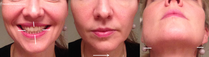
The picture confirms that you have a mandibular deviation to the left. On the smile view, we note that occlusal plane of the front teeth is canted to the right. The upper right teeth have more display when smiling compare to the left teeth. We see gums on the upper right side but not on the left side. The maxillary midline is inclined to the left while the lower midline is inclined to the right. Your case is very comparable to the
case depicted above. You chin is allso canted to the right. On the picture on the far right, we can see that right mandibular gonial angle is lower that the left mandibular gonial angle.
You say that this asymmetry begun to be noticeable in your late twenties and your are 40 years old.
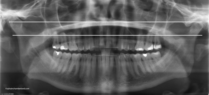
You definitely have after-effect of a hypercondyle, slowly but continuuously growing condyle. The maxilla has adapted over the years to the overgrowing condyle and skeletal and dental compensation occurred. You said that the radiography show a 15 mm increase of height of the ramus on the right side.
You had a bone scan in march 2014 that confirm an increase uptake of the right condyle. You are schedule for another bone scan next march 2015.
I agree with the approach of 2 bone scan, but i would have made them 6 months apart, not 12 months. Since march is coming soon, there no reason to not do another bone scan and if you can have your rendez-vous earlier, it is nice.
I understand that your asymmetric face affect your self esteem and despite this asymmetry you have a very good looking (vous êtes jolie). But because you are "jolie", i understand that you would like to correct the asymmetry of you face.
I ask you for a panorammic xray, which you will send me by next week i hope. I want to see if your condyle is elongated or enlarge. There is a slight difference. An enlarge condyle might indicate an osteochondroma (see slide 79 to 87 of the above keynotes). As Dr Bill Proffit said in a personnal communcation: "an elongated condylar process is more likely to stop growing than an enlarge condyle" (slide #87).
You asked me if it possible to prevent further growth. The answer is yes. A surgeon can do a high condylectomy of the growing condyle by shaving the cartilage surface. This is also describe in the keynote. A high condylectomy would be indicated if the 2
nd bone scan is positive. I would not recommend you to wait until the process of overgrowth is burned out because it might be active since the last 10 years and you are already affected with the asymmetry.
So let's assume that the bone scan is positive, i would consider a high condylectomy to stop the growth process and let you heal for 6 to 12 months. Meanwhile, i would start a comprehensive orthodontic treatment ot decompensate the dentition and plan for a bimaxillary surgery.

If the bone scan is negative, you could plan a comprehensive orthodontic orthognathic treatment plan and do the same type of surgery describe above.
Of course, the pro and cons of surgical approach will need to be discuss with you, the surgeon and the orthodontist.
WHat I wrote is an opinion that can not be consider a final diagnosis or a final treatment plan.
I have recommend you
Dr Martin Gaboury, a surgeon that graduated from the oral and maxillifacial surgery program at Hôpital Enfant-Jesus in Quebec. He is actually doing a fellowship in Bruges with
Professor Gwen R.J. Swennen.
You should get a rendez-vous with Professor Swennen and mention that you would like Dr Gaboury to assist during the consultation.
Gwen R.J. Swennen, MD, DMD, PhD, FEBOMFS, MSc
Professor, Division of Maxillo-Facial and Facial Plastic Surgery, Department of Surgery,
Bruges Cleft and Craniofacial Centre,
Three-Dimensional Facial Imaging Research Group (3-D FIRG),
General Hospital St-Jan Bruges,
Ruddershove 10, Bruges 8000, Belgium
Telephone: +32 50 453018
Best regards
Keep me inform.
Dr Sylvain Chamberland
.