The comments

Dr Sylvain Chamberland, Orthodontiste says:

Thank you Dr Sajeva for your questions.
1- Patients enrolled: All patients had a cl I occlusion and had their orthodontic treatment completed or within 2-3 months of completion. This is true for the 3 experimental groups.

2- There was no other skeletal deformity than vertical excess at the symphysis or lack of AP chin projection or both.

3-Hyperdivergent? The mean FMA was 34,06° (gr 1), 32,46° (gr 2), 34,76° (gr 3), 31,97° (Gr control). There was no statistical difference between groups regard FMA.

4- Unfavourable growth pattern.

This is answered in the discussion:  "Does repositioning the chin have a deleterious effect on mandibular growth? That is a valid concern and has been a major reason for delaying it until growth is essentially completed...In a growing individual with an indication for forward-upward genioplasty, data from our control group show that lip incompetency persists, facial convexity is maintained, bone resorption occurs at point B, and symphysis thickness has a tendency to decrease.
Our data show that the growth pattern do not change after a genioplasty, ie a vertical growth pattern do not become an horizontal growth pattern.

5- Repropose genioplasty.

No patient had to undergo a second genioplasty. However, if a patient have juvenile rhumatoid arthritis and has condylar resorption, he might benefit from an early genioplasty and a revision when growth is completed if a 2nd surgical phase is indicated.

6- Case selection: case selection was rigorous.

"For all subjects, the recommendation for genioplasty was based on clinical evaluation of the prominence and vertical position of the soft tissue chin relative to the lips and midface. Cephalometric data for pretreatment a-p chin deficiency relative to the lower incisors, the vertical distance from the incisors to the bottom of the chin, and the mandibular plane angle are shown in Table 1."

Warm regards

Dr Sylvain Chamberland

Dario Sajeva says:

The rationale of your study is interesting as well as all the interceptive surgery in growing patients.
1-Which patients have enrolled in the first group?
2-They were just "orthodontics" or also patients with skeletal deformities?
3-In the latter case have you enrolled surgical patients with hyperdivergent mandibular growth pattern ?
4-What has happened, in this case, with an clockwise unfavorable residual growth of the jaw?
5-it 'was necessary to repropose the genioplasty?
6-I think the selection of candidates should be rigorous and based on growth perspectives.
I wished to know your opinion an experience in this sense .
Greetings

Dr Sylvain Chamberland, Orthodontiste says:

If there is only one impacted canine I use the same appliance design, but with one cantilever spring.

I found an exemple of left impacted canine and a right fully erupted canine that is under treatment actually. It represent the mirror view of your case.

Here is the radiographic view of a deep palatally impacted left canine in a young adult.

Removable-appliance_cantilever-spring-for-impacted-canine-Chamberland-Orthodontiste-à-Quebec-M-A-Mor
The radiographic view show the direction of the force applied to the palatally impacted canine.

The advantage of a removable appliance is maximal anchorage and minimal side effect on neighboring teeth.

Occlusal-view-removable-appliance_cantilever-spring-for-impacted-canine-Chamberland-Orthodontiste-à-Quebec-M-AMor
Once the canine is visible into the mouth, fixed applaince is used to pull and place the canine into the arch and align in the 3 planes of space.

I hope this answer your question.

Best regards

.


Htet Naing says:

Please just may I know
Only for the removable appliance design that can solve the maxillary right permanent canine
But,with maxillary left permanent canine is fully erupted into the arch.
Just for one and please why do you chose these ?

Dr Sylvain Chamberland, Orthodontiste says:

I would like to show you another class II div 1 case that have IMPA at 104°.

Because of the proclined lower incisors, I distalized upper molars with a pendulum appliance and leveled the mandibular arch with intrusive arch mechanics "à la Burstone".

The case name is Roxane form slide 12 to 22.


Best regards


Dr Sylvain Chamberland

Dr Sylvain Chamberland, Orthodontiste says:

Hi Dr Medhat,

I understand that you may have difficulty with french language but believe me, french belgium orthodontist might have diffuculty with my "french canadian" accent as welll...HiHiHi!

I don't have plans to lecture to the Dutch part of Belgium, because I don't know that many flamish, except may be Dr Luc Dermaut that i met in Lebanon in 2005 during a scientific meeting.

However, i would certainly accept an invitation to lecture in english to the Dutch speaking orthodontist. Therefore, if you attend to the lecture in january, it will be easier to promote an invitation.

Please note that I lecture in english most of the time.

Here is the list of topic that will be presented:

1- Hemimandibular hyperplasia and facial asymmetry (75 minutes)


2- Idiopatic condylar resorption and arthrosis of the joint (75 minutes)


3- Functional genioplasty growing patients (45 minutes)


4- Fixed appliance management of class II malocclusion. SUS2 Corrector


5- Multidisciplinary treatment (Prostho, perio, orthognatic surgery, sleep apnea)


Best regards

Medhat says:

Thanks for your time and effort answering the questions.
I will try to attend your lecture in januari, however I guess it will be in French, which is not my strongest language.
Any plans giving lectures in the Dutch speaking part of Belgium (Flamish) ?

Dr Sylvain Chamberland, Orthodontiste says:

Thank you for your questions,

1- AP position of the lower incisors


If the lower incisors are at 100° to mandibular plane (IMPA ≥ 100°), chances are that there is a dentoalveolar protrusion and teeth need to be uprighted during treatment. Therefore, I may include extraction (4s/ 5s, 4s/4s or 5s/5s) in the treatment plan with the use of class II correction device.

Class II division 1 bimaxillary protrusion-Dr Chamberland orthodontist in Quebec City

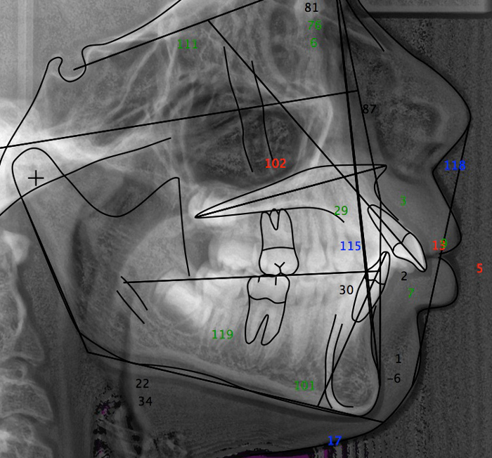
The ceph above show such a case with IMPA at 101° and bimaxillary protrusion and lip incompetency at repose. A fixed functionall appliance will be use to correct the class II relationship, but at the same time, lower teeth need to uprighted and upper teeth need to be retracted.

Class II division 1-Dr Chamberland orthodontist in Quebec City

This is the intraoral view of this case. You will notice crwding in the lower arch that will procline even more the incisors if aligment is attempted non extraction.

Therefore extraction was unavoidable. The ideal extraction plan would have been 4s/5s. However, a significant hypocalcification of tooth #25 made me consider extraction of upper 5s instead of upper 4s. The parents were advise that it will be more difficult to correct the class II relationship.

So for me, it is not a matter of dogma. It is a matter of treatment plan. Where do you want to place the lower incisors? If the answer is that you will accept their proclination, you may not extract. If the answer is that you want to upright them, then you shall extract.

This is the visual treamtent plan of the above case. You will note that the lower incisors are more upright and the upper incisors are retracted.

Visual treatment plan-Dr Chamberland orthodontist in Quebec City

A similar case is describe on slide 22 and 23 of the keynote Fixed appliance management of Class II correction.

2- Contraindication to Class II correction device


Crowding is not a contraindication to class II correction device if you have a solution to resolve crowding. This solution may be extraction of teeth. Class II correction device is not the only way to correct a class II. Maxillary molar distallisation via a pendulum or TADs may be an alternative solution. A class II hyperdivergent with vertical maxillary excess may be a contraindication for class II correction device.

In the above mentionned keynote, such a case is describe from slide 74 to 84. You will notice that I used TADS to intrude posterior teeth (upper and lower) and it was helpful to created a forward rotation of the mandible and to decrease face height. Once this was achieved, I use SUS2 class II correctors to finish into class I.

3- Loop in the lower arch


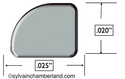
20x25 SS wire-Dr Chamberland orthodontist in Quebec CityI use SPEED™ .022 bracket slot. Therefore, the wire is .020 x.025 SS wire. It has a rounded edge. This may explain why it looks like a round wire, but is is not.

A .017 x .022 SS wire does exist for .018 bracket slot user.

To create this loop, I use an tweed omega loop plier (Dentronix D236).

.

Since you mention that you are a collegue from Belgium, please note that I will lecture for the Société Belge d'Orthodontie le 29 janvier 2015 à l'Hôtel Métropole, Place Bouckère.

Please come and introduce yourself.

Medhat says:

Dear Dr.Chamberland,

1) Do you consider using forsus or any other fixed functional appliance if the lower incisors to the mandibular plane angle 100° or more? Do you respect the 95° angle dogma?

2) Do you suggest certain contraindications for forsus? eg. crowding, gummy smile?

3) is the loop in the mand. Arch bent in a .018 stst wire? It seems like round wire from the pictures.

Thank you in advance,

College from Belgium

Al Bishop says:

Hi Sylvain.
I love your blog, honesty and the research that you share with people. I would like to get to know you.

Al

Dr Sylvain Chamberland, Orthodontiste says:

Yes, it is normal. It may be blood clot and granulation tissue. This tissue may cover the exposed canine.

Kasey says:

I had expose and bond procedure with a open hole in the roof of my mouth. Little over a week out and the hole has filled with hard dark tissue. Is this normal

Dr Sylvain Chamberland, Orthodontiste says:

Dental trauma on primary teeth

Hi Dr Templo,

I agree that it may not be necessaury to suture the frenum and let it heal by itself. The accident occurred a week ago and it would be too late. At this age, healing is often very fast and there will be no sequelae for the frenum.

Concerning the tooth buds, it would be important to know if the primary teeth were hitted directly, if they were mobile, if they were displaced, if there was bleeding.

Here are some possible diagnostics:
Concussion:

An injury to the tooth-supporting structures without increased mobility or displacement of the tooth, but with pain to percussion and without gingival bleeding.
Subluxation:

An injury to the tooth supporting structures resulting in increased mobility and pain to percussion, but without displacement of the tooth. Bleeding from the gingival sulcus is evident if the child is seen shortly after the accident.
Extrusion:

Bo-Elod-iof-traumatisme-dentoalveolaire-dent-primaire-extrusion-Chamberland-Orthodontiste-a-Quebec-21-08-2013Partial displacement of the tooth out of its socket
An injury to the tooth characterized by partial or total separation of the periodontal ligament resulting in loosening and displacement of the tooth. The alveolar socket bone remains intact. In addition to axial displacement, the tooth usually will have some protrusive or retrusive orientation.

In the picture on the right, white arrow show the extruded primary teeth. The black arrow show site of bleeding resulting from separation of the periodontal ligament.

If you need more information, I recommend that you visit the website The Dental Trauma Guide. It contains all the info you would need.

Dr. Francy Templo, DMD says:

Dear Dr. Sylvian Chamberland,
I have a 4 year old girl patient who came for consultation of her frenum accidentally cut crosswise upon her fall on the stairs, hitting the upper anterior and labial frenum. She had her antiobiotic therapy and looks normal on her smiling profile but she is just complaining of some sort of pain when such part is being touched obviously because the accident happened just a week ago. The parents are praying for a normal healing process so she will not undergo suturing. May I asked for your opinion if that could be possible that it shall heal by itself? I am just worried of the developing tooth buds underneath if disrupted of its normal growth which might cause an abnormality in its eruption and which might call for orthodontic concern in the future. Please advice. Thank you.

Myriah Phillips says:

Thank you for explaining these issues in such a way that anyone could understand. Hats of to you.

Dr Sylvain Chamberland, Orthodontiste says:

Thank you for your kind remarks.


I was able to log the link you provided me. I agree that your son have a rare issue of external resorption at the tip of the crown (cuspid tip). The case of your son is very particular.The prognosis is not very good.

Dr Sylvain Chamberland, Orthodontiste says:

No. It is not necessary to use open the bite while doing maxillary protraction. As the maxilla is pulled forward and down, relative extrusion occur posteriorly and correct the deepbite.

yemi says:

very interesting results. was there a need to prop open the occlusion anteriorly, with maybe a posterior bite plane in any of the cases?

Birgitta Bower says:

I just found your website! Which is astonishing, since I've been delving into impacted canines now for 2 years and have created a website
for the benefit of other parents and to document my son's case.

We live in the San Franciso Bay Area, but went to Jerusalem in March to see Dr. Adrian Becker and have my son's canines exposed for a third time. My son's canines happen to also be afflicted with PEIR (preeruptive intracoronal invasive resorption), even Dr. Becker has never seen a similar case. I have posted his thoughts, hypothesis and his handling of my son's case on the website if you are interested.

I have found that it is a very painful process to try to educate yourself as a parent to be able to find the right option for your child. A resource like your website though is a true godsend and I thank you on behalf of all parents in this position.