The comments

Dr Sylvain Chamberland, Orthodontiste says:

Thank you for your kind remarks and I am glad that this blog help you.


Please note that the french version of this page contain more photos en comment. You may be interested to look. Even if you can't read french you will get interesting info.


Best regards


Dr Sylvain Chamberland

Mel says:

This was very informative and alleviated any doubt regarding my decision to not put my 6 year old through this procedure.
We were referred to a surgeon for a consult, because her doctor was concerned that the frenum would cause her central incisors to erupt in a way that would prevent them from closing.
After researching and reading up on the techniques, results and both sides of the debate, I just don't think we need to put her through this, yet. As stated here and elsewhere, we'll just wait til her canines erupt and take it from this.
Honestly, before her doctor brought it up, I had never heard of a frenectomy or frenotomy. It seems to be much more common than I initially thought.
Thank you for providing such thorough information and pictures.

Terry D. Carlyle says:

Sylvain
As always a great job on these issues of concern to the public and the profession.
Keep up the good work.
TDC

Dr Sylvain Chamberland says:

No. It is not possible.


If the patient really have a cl II div 2 relationship, lingually inclined central incisors and deep overbite and your ceph tracing show ANB angle < 2°, chances are that you mistraced the ceph. You misplaced either Sella, A point or B point.


If the patient really have ANB < 2°, he could have lingualy inclined central incisors that created an anterior crossbite. If this is the case, chances are that your diagnostic is wrong. It is not a cl II div 2 but rather a pseudo class I or a class III. You may also be reading the molar relationship on primary 2nd molars instead of permanent 1st molars.


That is what my 18 years of experience of teaching orthodontics to undergraduate student told me.
Thank you for asking.


Best regards

sylvia says:

sorry i have a question,
is it possible that class II division 2 have skeletal class III in cephalometri? (SNA-SNB <2)
i'm dentistry student and find my 10 years old patient having this issue. please help :(

Dr Sylvain Chamberland says:

I am happy to learn that you are going ahead with a solution that seems OK to me. I would like to reassure you about the central incisors that have resorbed. Know that as soon as the canines are no longer near them and stop putting pressure on them, the resorption will stop. So even if 3, 4, 7 mm of roots have resorbed from the central incisors, once the treatment is over, this does not mean that these teeth have a worse long-term prognosis. She can live with this all of her life like we say back here.


I imagine that your orthodontist has put appliances in the mandibular arch to match both dental arches. Otherwise, there is another problem that will get to you sooner than later. You wrote on 8 December that she had a Class II relationship. This is compatible with an orthodontic treatment where 2 teeth in the maxillary arch are extracted (the lateral incisors in Lorène's case).


Canine lateral substitution and Class II molar relationship-Dr Chamberland orthodontist in Quebec City

Canine lateral substitution and Class II molar relationship.


I show you an example of a treatment where the missing lateral incisors were substituted by canines and there is a Class II molar relationship. There were no extractions in the mandibular arch.

Eric says:

Hello Doctor Chamberland,

Here are news on Lorène.

This time, this is it... The orthodontist will try to go get the canines. It is a real art... He will first remove the 2 external incisors (which will anyway fall out in the short term since the roots are damaged). Then, to go get the impacted canine, he will move the incisors on the side and pull on it. He will try to save at best the main incisors (but they are, according to him, also doomed in the medium term, because they have also resorbed to a lesser degree and one more than the other. We will see...) The canine which is in the palate will be pulled backward in the meantime. He will then put everything back where it belongs... It is complex but the goal is to save the canines which are healthy teeth while the incisors are doomed in the medium and short terms. Lorène should undergo two surgeries under local anesthesia.

This seems to me like the best that we can do. I am happy to have stopped beating about the bush and a little calmed down even if we are in for a heavy treatment. Thank you for your advice.

Regards,

Eric Lacasse

Dr Sylvain Chamberland says:

Madame Caroline,


Facial-asymmetry-_-Cant-of-the-occlusal-plane-Chamberland-Orthodontiste-QuebecYour 23 years old daughter has a facial asymmetry to the left with a vertical component, canting the occlusal plane to the right like this young girl on the left pict. The girl, in this exemple had a bone scan and the intake of the right side was the double of the left side (3,07 versus 1,56).


We could have decide to wait and redo a bone scan in 6 months to see if the hypergrowth of the condyle is burning out, but since the asymmetry was evident, the question was: how much more facial asymmetry is she willing to tolerate before growth phaseout?


Therefore, the decision was to do a high condylectomy as soon as possible. This case is shown in the keynote presentation above.


One have to remember that significant facial asymmetry usually need bimaxillary surgery. This was the case of this girl, but she declined further treatment and the high condylectomy stopped asymmetric facial growth.


In the situation of your daughter, i doubt it would be an undiagnose condylar fracture at an early age even if it is not impossible.


If you could send me a similar photograph of your daughter and a panoramic radiograph, I could provide more specific information. You know my email. 


Thank you Caroline for this excellent question. I value your concern about your daughter.


Best regards


caroline says:

I have read Dr Chamberland's report on facial asymmetry which was very helpful in the case of my daughter who is 23 and has increasing facial deformity, which is now very evident due to accelerated growth over the last 18 mths.
She has significant mandibular asymmetry, mainly on the vertical dimension and significant cant of her maxilla, down on the right side.
She has had a technetium scan and there is a very increased uptake in the right condylar area, growth is still active.
OPT radiograph shows an element of hemimandibular hypertrophy with increased body height on the right side.
We have been told that no treatment can be started until the growth has burned out.
I would appreciate a second opinion as at the age of 23, my daughter is very aware and upset regarding her change of appearance of which no evidence was given to the length of time for growth to stop or other alternatives. She refuses any photographs to be taken front on, which is affecting her confidence.
I am not a profession person in the medical world and would deeply appreciate a second opinion and whether prevention of further growth or exision of bone at the head of condyle, removing the condyle head, etc, can be done as well as a sagittal split osteotomy before growth stops.
As examinations were done in November 2013, you may well advise a 2nd bone scan in April to assess growth and earlier trauma as Dr Chamberland highlighted and further advise would be gratefully accepted.

Dr Jules E. Lemay III, orthodontiste says:

A CBCT is a lot more serious to help in diagnosing an impacted canine and its relationship with nearby structures. To see examples where this imaging technique was a precious diagnostic help; follow this link

Dr Yves Gagnon, prosthodontist says:

Congratulations Dr Chamberland for this exhaustive reply but nonetheless very clear on several points of view.

I believed for several years that occlusion was a major factor in TMDs. I thus can have some empathy for all these dentists who believe this myth a lot.
It is a little sad, of course, to see that some of us become such fanatics that they even plan conferences and invite physicians, physiotherapists, chiropractors, radiologists, etc., etc., etc. Even there, I can understand.

Unfortunately, these different healthcare professionals are rarely aware of the importance of the scale of scientific evidence, even less of the different levels of evidences and what else to say about their capacity to criticize objectively a conference on dental occlusion while they know nothing about dentistry???

Nevertheless, it is possible that the speaker himself is aware of this ignorance and takes advantage of it by using logic, mythical beliefs, numbers to orchestrate everything well with some speaker charisma to "catch" disciples, making it easier to enlarge the group and provide additional support to the so-called philosophy (open question).
Unfortunately, if we do not want to lose our profession and bring it to the level of alternative medicines, we must object to all of these philosophers of occlusion in great numbers.
Having completed several of these philosophies of occlusion (Skavicek, Dawson, neuromuscular) myself, I was fortunate to become confused enough to go back to school and understand the importance of this famous scale of scientific evidence. I believe that the recent report released by the ODQ is an excellent first step in the right direction; unfortunately, I notice through my training seminars that there are still just a few rare dentists who know about this report. It is still an excellent initiative from the ODQ to protect the public against these PHILOSOPHERS.

Some of the dentists who register for my training seminars also (unfortunately) have taken a lot of these training seminars. Kudos to those who, despite dozens of thousands of dollars invested, still had the courage to come back on the right path (the one guided by the scale rather than mythical beliefs of a GURU).

We thus still have a lot of work to do but more and more dentists are aware of this scale. Several new graduates are aware of this scale and they will be a lot harder to recruit, "catch", be convinced by GURUS.

Let's continue to work in that sense, our profession will benefit from it in the end.

Dr Yves Gagnon, prosthodontist.

Dr Sylvain Chamberland says:

Good morning Dre Roy,


I apologize for taking so long to answer. I was busy in the last few days.


I take note of your comment about "preconceived ideas". An exhaustive research published in the Cochrane library was precisely performed to find an answer to the preconceived idea that occlusal adjustments can treat or prevent symptoms or temporomandibular disorders.


But let's first differentiate two concepts:


1-Experience-based view.


Clinical experience and years of experience are the only important things for the practitioner. Usefulness of science is often denied. Anecdotal evidences are sufficient to make a clinical decision and are better than science. TMD diagnosis is based on meticulous analysis of occlusion. Recommendation is to use occlusion protected by the canine. Any position of the condyle other than supero-antero-medial position causes TMD. The use of articulators in orthodontics is favored.


2-Evidence-based view.


Science and scientific method are important to the practitioner. Benefit and usefulness of science are proven. Testimonies, case studies and non-objective experience are inadequate or insufficient to make a clinical decision. No definitive tests exist in TMD diagnosis. The gold standard for diagnosis is based on case story, clinical examination and TMJ imaging when indicated. Occlusion is not the main cause of TMD, but possibly plays a minor role in its etiology. TMD are a whole, a collection of disorders. The position of the condyle per se is not directly associated with TMD. Biological concept of a functional occlusion includes all kinds of occlusion (group function or canine guidance) but not occlusal interferences (balancing and protusion contacts are tolerated). Articulators are not necessary in orthodontics.


Rinchuse DJ, and Kandasamy S. Evidence-based versus experience based views on occlusion and TMD. AJODO 2005, feb;127(2):249-54


I would like to point out that even if I am a practitioner cumulating 30 years of experience, I am part of those who believe in evidence-based dentistry.


Hierarchy of quality of scientific evidence
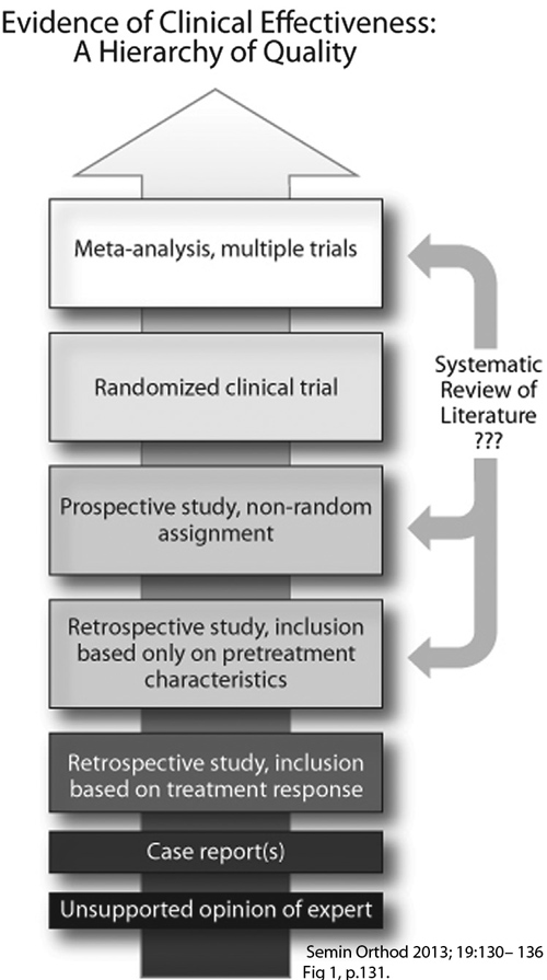

Hierarchy of scientific evidences-Dr Chamberland orthodontist in Quebec City Drs Peter Dawson and Mark Piper's researches and work have to be placed, like it or not, in the "unsupported opinion of expert" category, at the bottom of the scale of scientific evidence. Holy Koh and Peter Robinson's article on occlusal adjustments from the Cochrane library is located at the top of the scale along with meta-analysis including multiple trials. I invite you to read the whole article: Occlusal adjustment for treating and preventing temporomandibular joint disorders (Review)


It is interesting to note that none of Drs Dawson and Piper's research work was selected in this meta-analysis. It is likely to believe that their work didn't meet the high selection criteria and absence of bias to be considered and included in the meta-analysis.


.


Mark Piper and "Piperism"


A very good example of preconceived idea is provided to us by Dr Mark Piper when he describes TMJ avascular necrosis by saying that compression of the TMJ perforating blood vessels occurred. He tries to link avascular necrosis caused by a fracture and collapse of the femoral head where a main artery and branches of lateral and medial circumflex femoral artery are located.
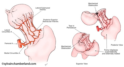

Femoral artery-Dr Chamberland orthodontist in Quebec City
TMJ Piper-Dr Chamberland orthodontist in Quebec City

He pretends that anterior disc displacement compresses “so-called” TMJ perforating blood vessels and causes avascular necrosis of the condyle.


But this artery does not exist.


Piper uses avascular necrosis concept to promote his microsurgery technique where he perforates the head of the condyle to promote vascularization.
He showed histological bone cuts that he removed during these perforations while saying that "his pathologists" said that it was "dead" bone.
But when these samples were re-analyzed by independent and renowned pathologists, they all showed that the bone was alive and they refuted his whole avascular necrosis concept, which scientifically put an end to the discussion.
Piper's results concerning the use of adipose tissue grafts to treat TMJ internal derangements and condylar resorption were never published in any renowned journal where articles are revised by peers before publication (peer-review journal). To this day, nobody has published an article showing that someone had been able to reproduce Piper's results.

Definition of reproducibility in science


Reproducibility is the degree of similarity between measures or observations when reproducing a procedure, an experience, an analysis by different people in different places (hospitals, universities, countries). Reproducibility is part of the precision of the tested method.


Arterial blood supply of the TMJ
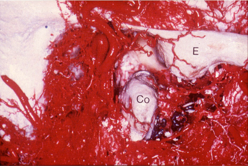


TMJ arterial blood supply Artery LMercury-Dr Chamberland orthodontist in Quebec City

Arterial blood supply of the TMJ comes from multiple sources (external pterygoid muscle, cancellous bone, internal pterygoid muscle, posterior attachment vessels and transverse facial artery). Anybody having performed a surgery on a TMJ knows that blood supply comes from multiple sources. See the attached picture showing the arterial network and see Dr Carl Bouchard’s video clip while he performs a costochondral graft on one of my patients. Notice the hemorrhage when he unfortunately cuts a small artery when working on the left side (CCG link).


The Québec Association of Orthodontists recently invited Dr Alain Aubé to hear his point of view on TMJ. Let’s say that several colleagues from the Association expressed huge hesitations against the concepts conveyed by Dr Aubé. Numerous sophisms were noticed. We witnessed a display of preconceived ideas like we had rarely seen in a continuing education course.


We do not deny the use of magnetic resonance imaging in some TMJ pathologies, such as disc displacement, but diagnosis of TMD cannot be limited to magnetic resonance imaging.


Treatments of TMD have to rely on scientific basis.


Conclusion


The mistake to avoid is to put occlusion at the center of all TMJ problems and believe that the solution to TMJ problems is through only one functional occlusion model with canine guidance and bilateral contacts equally adjusted to 8-micron shimstocks.


Prevalence of Class I, Class II or Class III malocclusions is about equally distributed between men and women. Dismorphism is not based on gender.


Differences in types of malocclusions (Class I, Class II or Class III) exist depending on the population observed: white (Caucasian), Afro-American, Asian or Hispanic.


Incidence of TMD in the population in general is of 2 women for one man.


Incidence of patients suffering from TMD is of 10 women for one man.


Dr Larry Wolford (Atlas Oral Maxfacial Surgery Clin N Am 19 (2011) 243-270), in a research comprising 1369 consecutive patients from 8 to 76 years of age referred for TMD, observed a proportion of 78% of women and 22% of men. 69% of affected patients mention that their problem started in their teenage years. He concludes that TMD mainly develop in teenage girls.


We can think about the following:


If occlusion were the cause, it is likely to pretend that the M-F distribution would be more balanced.


In this context, isn't it unlikely to pretend that occlusal adjustment prevents TMD knowing that women are more affected by this kind of problem and that there are no differences between men and women in prevalence of malocclusions?


In conclusion, I will quote Dr Louis Mercuri:


"It is essential that TMJ osteoarthritis be presented as the pathologic entity it is in the same terms as our colleagues discuss osteoarthritis in orthopaedic circles.


To not do this only exacerbates the problem that everyone dealing with this entity patients, clinicians, insurance carriers, and so forth has with TMJ osteoarthritis, because they do not consider it as the orthopaedic (medical) pathology that it is, but rather a purely dental TMJ problem."


L.G. Mercuri Oral Max Surg Clin N Am 20 (2008) 169-183


Finally, I recommend to you the excellent book from Drs Charles Greene and Daniel Laskin which was just released to rectify a lot of your preconceived ideas that were transmitted to you by Drs Dawson and Piper.


Treatment of TMDs:
Bridging the gap between advances in research and clinical patient management
, ©2013 Quintescence Publishing Co.

Eric says:

As far as the lower arch is concerned, it is favorable, my daughter is in Class II.

Regards,

Eric

Dr Sylvain Chamberland says:

Mister Dufour, you are a practical guy. No fooling around.

Dr Sylvain Chamberland says:

I am very happy to learn that one of the canines has erupted by itself. I assume that it is the left one because its position seemed less problematic on the panogram.

A CBCT will allow defining the position of the impacted canine relatively to the roots of the incisors more precisely.

The fact that a canine is in the mouth changes everything. A different treatment plan can be considered. This treatment plan must include treatment of the mandibular arch, the likely lack of space in the maxillary arch and the fact that the roots of one or two incisors have resorbed to the right and the feasibility of bringing the impacted tooth into function.

I do not see any problems keeping both canines, but the right canine needs to be taken away from the incisors to stop the damage. This should have been done a long time ago. The problem is that there is no global approach for the maxillary arch as well as the mandibular arch.

A lot of time was wasted by a lack of understanding of the case and the inherent risks to the impacted canines.

Good luck

Kent Dufour says:

Homeopath???? I quite laughed. I am a mechanic and I am one of Dr Chamberland's patients. And there is no use in having a degree at university to know that a homeopath cannot do anything for your daughter’s teeth. Apart maybe from wasting your young daughter's time.
When I work with a new young mechanic and he tells me that he does not understand an electrical problem, I tell him that he can continue believing that it is magic, or consult the technical publications and understand the problem. On the same level, I do not believe that a phoney homeopath can move teeth...

Eric says:

Hello Doctor Chamberland,

We went to see the homeopath who recommended that we go see a colleague homeopath/dentist who just recommended to us to go for a cone beam computed tomography...

We will go next Wednesday...

In the meantime one of the canines came out in the palate, the other one is still hidden...

I will keep you informed,

Regards,

Eric

Valérie Roy says:

I invite you to look through Dr Peter E. Dawson's researches, who devoted his career to occlusion. His researches will maybe change preconceived ideas on the latter. Moreover, you will be able to consult Dr Mark Piper's work who, because of extensive assessment of TMJ MRI, explores TMD in a lot more details. Huge breakthroughs that are unfortunately still not well known.

Dr Sylvain Chamberland says:

Thank you Dr Proffit,


Your input is very much appreciated. In the AJODO 1980′s article, there is a picture of an « Hemi-Frankell » and I do remember having see a similar patient in an edition of Contemprary Orthodontics.


Happy Thanks Giving


Best regards


Sylvain